The femoral artery pulse is palpated and a vertical incision is made over the pulse.
In the case of inflow disease, a palpable pulse may not be present, and thus Doppler ultrasound or landmarks (midway between pubic symphysis and anterior superior iliac spine) provide a guide.
Once through the subcutaneous tissues, sharp dissection is used to isolate the common femoral artery proximally to inguinal ligament and distally past the superficial femoral artery to avoid thermal injury.
Branches of the common femoral artery arise medially and laterally. Therefore, dissection along the anterior surface of the artery avoids injury.
When dissecting close to the vessel, recall that spreading perpendicular to vessel will prevent inadvertent avulsion of small branches.
The superficial femoral artery, the common femoral artery, and the profunda are encircled with vessel loops prior to arteriotomy.
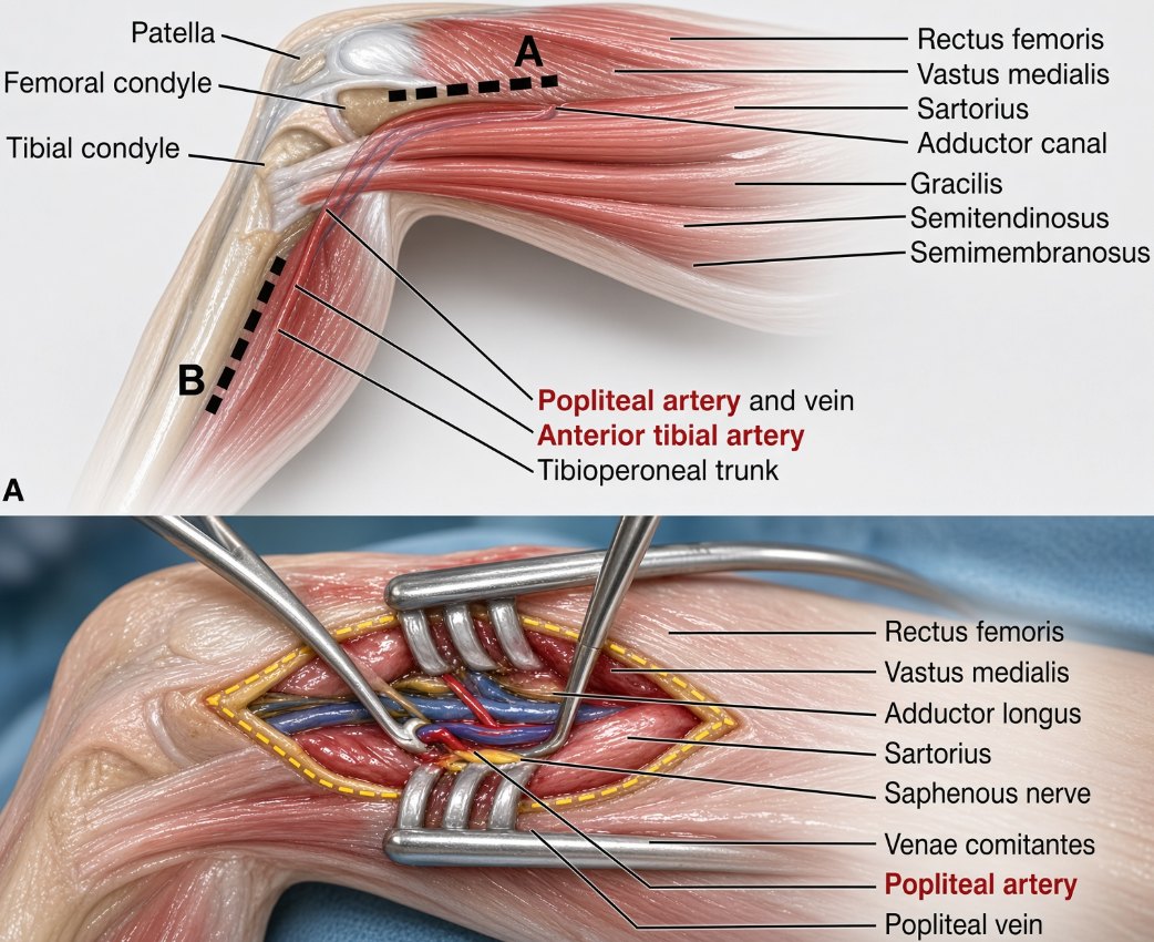
Above Knee Popliteal
This involves a longitudinal incision in the lower thigh, mobilization of the sartorius laterally, and incision of the fascia of the adductor magnus to enter Hunter canal and identify the artery.
Below Knee Popliteal
This involves longitudinal incision just medial to the tibia and division of the fascia of the gastrocnemius with mobilization inferiorly. Then it is possible to enter the popliteal fossa and isolate the artery.
Educational aid only. Verify every dose, indication, and guideline against current institutional protocol. This is not a substitute for clinical judgment.