Make a transverse incision two fingerbreadths above the sternal notch, and then open the platysma transversely.
Raise the subplatysmal flaps from the thyroid cartilage superiorly to the sternal notch inferiorly.
Retract the strap muscles laterally by longitudinally opening the midline raphe to expose the thyroid gland.
Exploration should begin in the location of a suspected abnormal gland (based on imaging) and then proceed to the ipsilateral gland, followed by the contralateral glands.
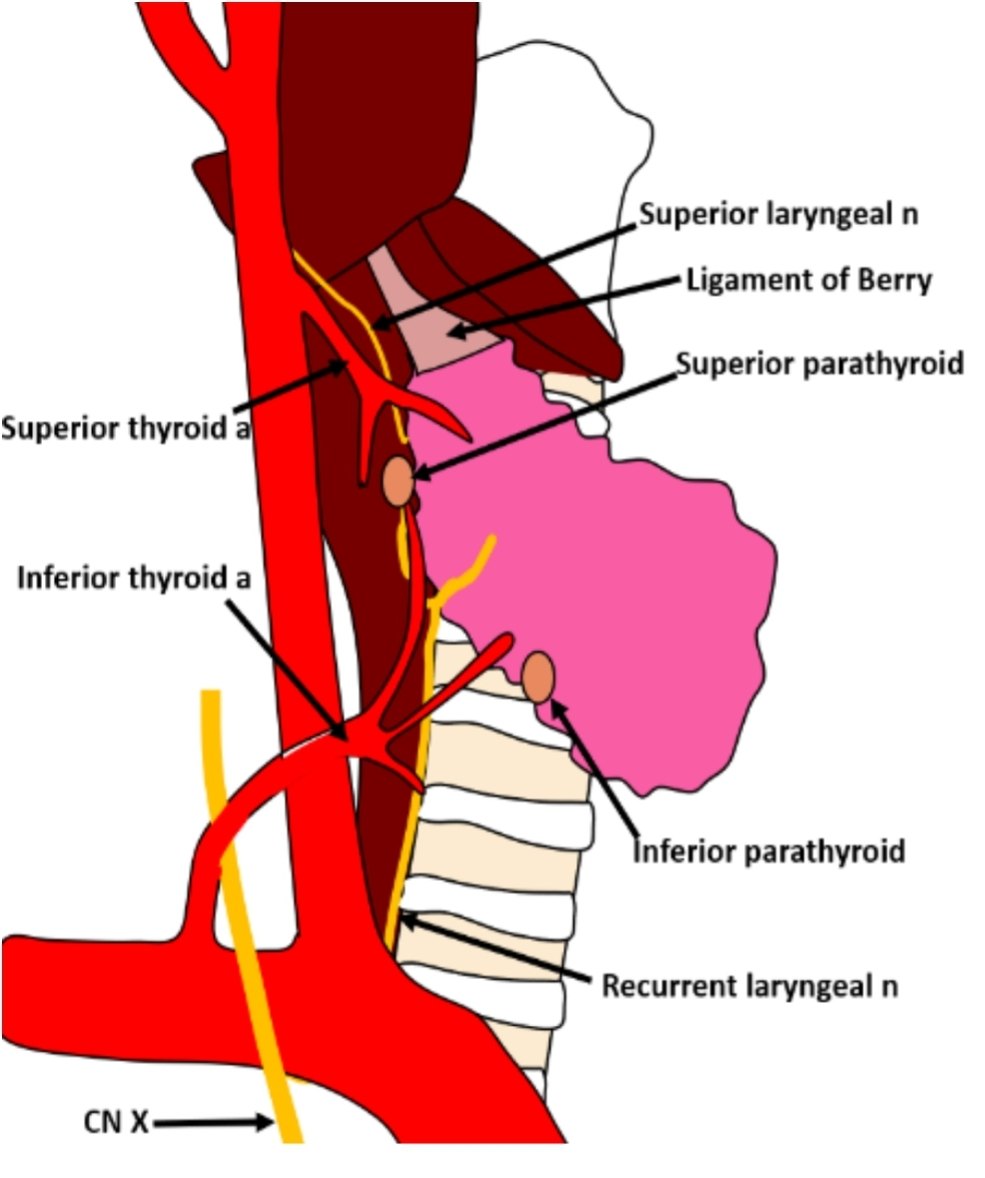
Gently retract the ipsilateral thyroid lobe medially to search the area around the thyroid gland and the tracheoesophageal groove for the parathyroid gland.
Division of the middle thyroid vein may facilitate visualization.
When an abnormal gland is identified, begin the dissection along the lateral aspect of the gland until the vascular pedicle is isolated.
Confirm the location of the recurrent laryngeal nerve prior to ligation of the pedicle.
Repeat the above steps to identify the remaining glands. Normal-appearing glands should not be resected.
If four-gland hyperplasia is suspected, a resection of three and a half glands will be necessary. Identify all glands before beginning resection. Leave 50 to 100 mg of the most normal-appearing gland in situ (preferably an inferior gland, which may be more readily accessed in the future). Mark this gland with a clip or stitch to facilitate localization if reoperation is required. Do this before resecting other glands to ensure the gland is viable.
Intraoperative nerve monitoring (IONM) may be considered, especially in the setting of reoperative surgery.
Current data regarding prevention of nerve injury with IONM are conflicting.
Educational aid only. Verify every dose, indication, and guideline against current institutional protocol. This is not a substitute for clinical judgment.