Usually have the patient in reverse Trendelenburg position.
Dissect the gastrohepatic ligament, identifying a possibly replaced left hepatic artery and/or accessory hepatic artery.
Identify the right crus and open the phrenoesophageal ligament.
Perform a blunt dissection of the crus off the esophagus anteriorly and circumferentially to the left crus (identify anterior vagus).
Divide the short gastric vessels to improve caudal dissection.
Create a posterior window behind the esophagus (identify posterior vagus).
Place a Penrose drain around upward retraction of the esophagus.
Make a crural closure, caudal to cephalad, interrupted, pledgeted, nonabsorbable suture at low pneumoperitoneum, leaving a 1.5-cm free space in the hiatus to reduce dysphagia.
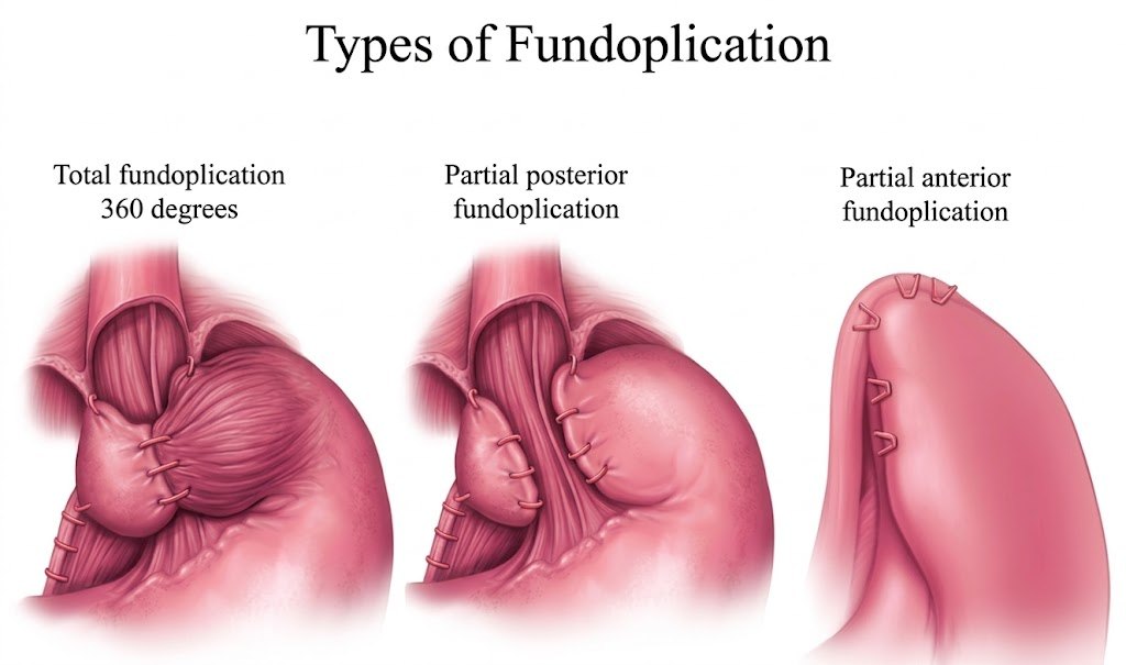
Perform fundoplication:
Bring the stomach fundus behind the esophagus and perform shoeshine maneuver.
Place a 56Fr or 60Fr bougie.
Perform a 360º wrap over the distal esophagus using three nonabsorbable pledgeted sutures, incorporating the esophageal wall.
Use esophagogastroduodenoscopy at the end to assess the adequacy of the wrap.
Educational aid only. Verify every dose, indication, and guideline against current institutional protocol. This is not a substitute for clinical judgment.